Introduction
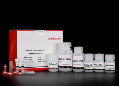
Magen has been manufacturing nucleic acid extraction systems for over ten years with a consistent focus on silica membrane purification technologies. The HiPure FFPE DNA Kit represents the column-based configuration within the Magen FFPE extraction portfolio and is designed for genomic DNA recovery from paraffin-embedded tissue sections.
The workflow combines optimized digestion chemistry and silica membrane adsorption to release fragmented DNA from formalin-fixed tissues while reducing inhibitor carryover during purification. Membrane structure and binding buffer composition have been refined through internal optimization cycles to support consistent DNA recovery from archived tumor samples.
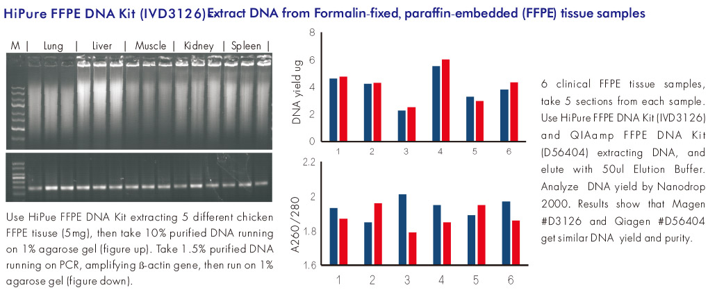
Internal comparison testing using FFPE tissues demonstrated stable DNA amplification performance during qPCR analysis under defined extraction conditions, with performance comparable to widely used column-based FFPE purification systems.
Laboratories operating different workflow formats may refer to:
HiPure FFPE DNA Kit functions as the Column reference model within the Magen FFPE extraction system.
Details
Workflow
Workflow Overview
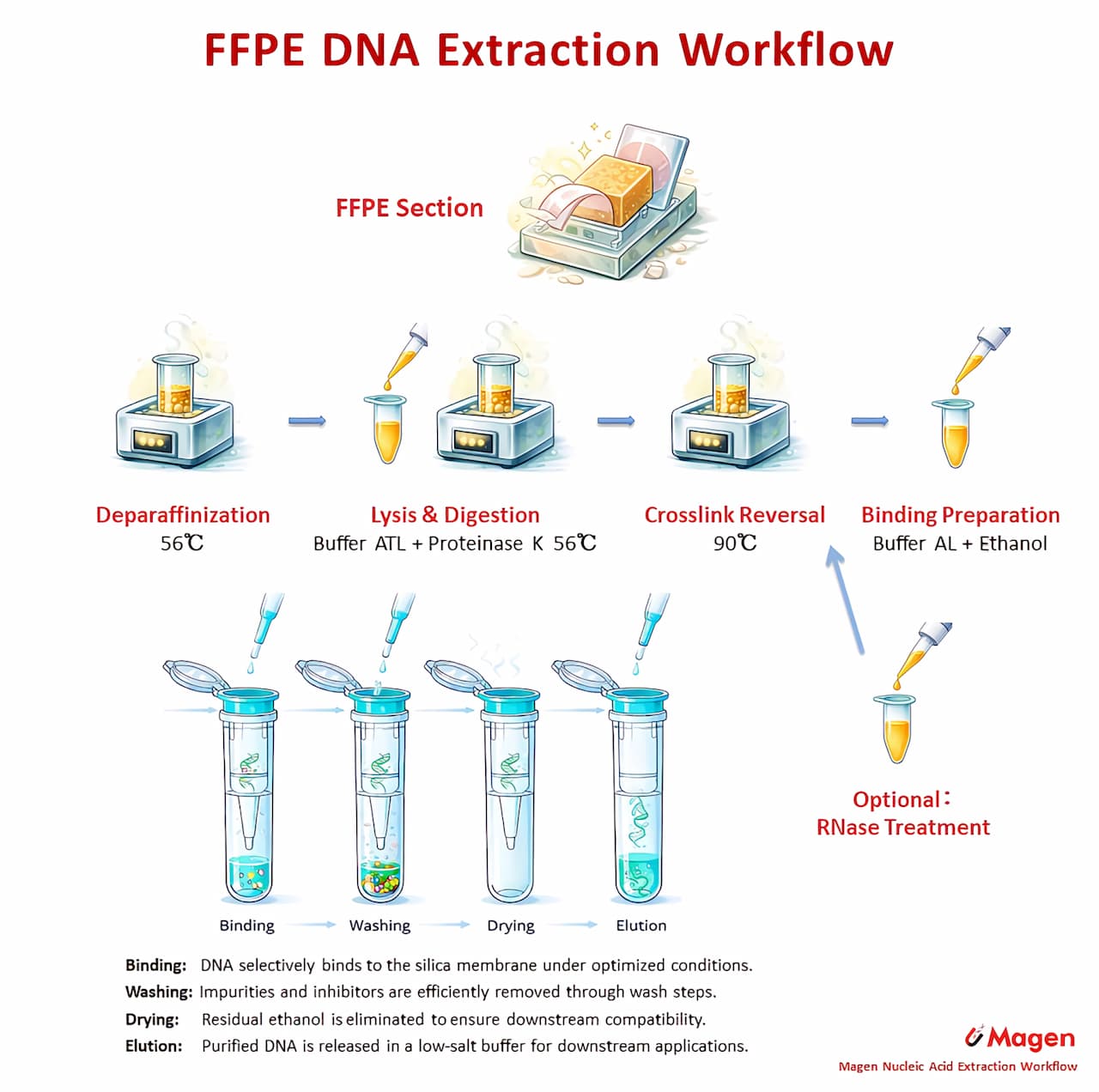
The HiPure FFPE DNA workflow uses a silica column–based route for purification of genomic DNA from FFPE sections. After deparaffinization, proteinase K digestion and heat-assisted crosslink reversal, DNA binding conditions are established and the released DNA is captured on the silica membrane. The workflow then proceeds through washing, drying and elution to recover purified FFPE DNA for downstream molecular analysis.
Sample Handling Logic
This workflow is designed around the main challenges of FFPE DNA recovery: paraffin removal, digestion completeness and reversal of fixation-related modification. The most sample-dependent variation is concentrated in the digestion stage, where tissue type, section thickness and block condition can influence processing time. Once DNA binding conditions are established, the downstream column purification steps remain consistent.
Time and Workflow Characteristics
Under typical manual operation, the overall workflow usually requires about 2.5–14 hours, mainly depending on FFPE tissue digestion requirements. For detailed step-by-step handling logic, workflow guidance and estimated processing times, please refer to the Workflow Note in the Download section.
Specifications
|
Features
|
Specifications
|
|
Main Functions
|
Isolation total DNA from FFPE tissue samples
|
|
Applications
|
PCR, southern blot and viral DNA detection, etc.
|
|
Purification method
|
Mini spin column
|
|
Purification technology
|
Silica technology
|
|
Process method
|
Manual (centrifugation or vacuum)
|
|
Sample type
|
Formalin-fixed, paraffin-embedded (FFPE) tissue and sections samples
|
|
Sample amount
|
<20mg
|
|
Elution volume
|
>15µl
|
|
Time per run
|
2.5~14 hours, depending on FFPE tissue digestion requirements
|
|
Liquid carrying volume per column
|
4ml
|
|
Binding yield of column
|
100μg
|
Engineering Characteristics
Crosslink Reversal Digestion
The lysis workflow facilitates controlled reversal of formaldehyde-induced crosslinks commonly present in FFPE tissues, improving accessibility of genomic DNA during extraction.
Silica Membrane DNA Adsorption
A multi-layer silica membrane enables adsorption of fragmented DNA typical of FFPE samples while maintaining predictable filtration behavior.
Inhibitor Removal
ChemistryOptimized wash buffers reduce residual paraffin components, protein fragments, and PCR inhibitors during purification.
Reproducible Column Performance
Membrane production and buffer formulation are monitored through internal QC procedures to maintain stable DNA recovery across production lots.
Technical Validation
DNA Amplification
PerformanceDNA extracted from FFPE tissue sections supported qPCR amplification across multiple template dilutions, confirming suitability for sensitive molecular detection workflows.
Comparative Extraction Evaluation
Internal experiments comparing FFPE purification workflows demonstrated consistent DNA recovery and amplification behavior relative to established column-based extraction systems.
Compatibility with Downstream Workflows
Purified DNA was compatible with sequencing preparation and mutation analysis workflows commonly used in oncology research laboratories.
Lot-to-Lot Stability
Extraction performance remained stable across validated production batches under internal verification testing.
Application Scenario Summary
FFPE DNA extraction workflows are selected according to the downstream assay. For focused PCR-based applications such as MSI-PCR, Sanger sequencing or mutation-specific analysis, the main requirement is amplifiable DNA from FFPE sections. For sequencing-oriented applications such as WES, targeted NGS, methylation profiling, HRD-related analysis, TMB evaluation or matched tissue-reference studies, fragment handling, inhibitor background and library preparation compatibility may become more important.
The selected examples below summarize published research workflows involving Magen FFPE DNA extraction formats, including D3126 / HiPure FFPE DNA Kit, D6323B / MagPure FFPE DNA Kit and D6323D / MagPure FFPE DNA Kit High Pure. These examples are organized to illustrate typical FFPE DNA application contexts rather than to compare kit performance directly.
|
Application Scenario
|
Related Format
|
Sample / cfDNA Source
|
Downstream Research Use
|
|
De novo high-grade meningioma TERT promoter mutation analysis for post-radiotherapy progression risk research
|
D3126
|
FFPE tumor tissue from de novo WHO grade 2–3 high-grade meningioma patients treated with postoperative radiotherapy
|
Genomic DNA extraction for PCR amplification and Sanger sequencing of TERT promoter mutations, supporting analysis of tumor progression, PFS and postoperative radiotherapy outcome
|
|
CRC and gastric cancer MSI-PCR panel comparison for optimized molecular pathology testing
|
D3126
|
FFPE biopsy and surgical specimens from colorectal and gastric cancer patients
|
Genomic DNA extraction for PCR-based MSI status detection using NCI, five-mononucleotide and six-mononucleotide panels, supporting comparison of panel performance, MSI-L interpretation and optimal MSI-PCR workflow selection
|
|
Pulmonary nodule malignancy assessment using paired FFPE tissue and plasma methylation profiling
|
D6323B
|
Malignant and benign FFPE lung tissue samples from patients with CT-detected pulmonary nodules
|
Tissue gDNA extraction for DNA methylation sequencing, paired tissue–plasma methylation concordance analysis and validation of an integrative blood-based model for distinguishing malignant lung nodules from benign lesions
|
View more application scenarios
|
Application Scenario
|
Related Format
|
Sample / cfDNA Source
|
Downstream Research Use
|
|
Pancreatic adenocarcinoma CNV-driven molecular subtyping for prognosis and treatment stratification research
|
D6323B
|
FFPE tumor samples from pathologically diagnosed pancreatic adenocarcinoma patients in a 608-patient Chinese cohort
|
Tumor genomic DNA extraction for 566-gene / 764-gene targeted sequencing, somatic mutation profiling, germline HRR variant analysis, CNV-based molecular subtyping and relapse-risk model construction
|
|
NSCLC postoperative MRD monitoring using tumor-informed personalized ctDNA analysis
|
D6323D
|
FFPE tumor tissue sections from early-stage NSCLC patients, paired with peripheral blood or buffy coat normal DNA
|
Tumor gDNA and matched normal gDNA extraction for WES, patient-specific PROPHET panel design, postoperative plasma ctDNA MRD monitoring, recurrence risk prediction and TNMB prognostic classification
|
|
FFPE tumor HRD assessment using genomic scar and allele-specific CNV analysis
|
D6323B
|
FFPE human tissue samples from ovarian and breast cancer cohorts
|
Tumor DNA extraction for capture-based HRD panel sequencing, allele-specific CNV analysis, Genomic Scar Score modeling and PARP inhibitor response prediction
|
|
Early TNBC HRR mutation profiling for immune infiltration and prognosis association research
|
D6323B
|
Archival FFPE tumor blocks from early triple-negative breast cancer patients
|
Tumor genomic DNA extraction for HRR-targeted NGS, mutation spectrum analysis, CD8+ T cell / PD-L1 association study and combined immune-genomic prognosis stratification
|
|
Pediatric hepatoblastoma trans-ancestry mutation landscape research using paired FFPE tumor and noncancerous tissue WES
|
D6323B
|
FFPE hepatoblastoma tumor samples and corresponding noncancerous liver tissues from pediatric patients
|
Tumor and matched noncancerous tissue DNA extraction for WES, somatic mutation detection, copy number alteration analysis, pathway enrichment and trans-ancestry genomic comparison
|
|
Matched FFPE tumor sequencing for distinguishing clonal hematopoiesis interference in liquid biopsy
|
D6323B
|
FFPE tumor tissue samples from Chinese pan-cancer patients with matched PBL DNA and plasma cfDNA
|
Tumor genomic DNA extraction for targeted sequencing, validation of candidate CH mutations, exclusion of tumor-derived variants and improved interpretation of cfDNA liquid biopsy results
|
|
LUAD tumor mutation burden and immune phenotype profiling for EGFR-mutant immunotherapy response research
|
D6323B
|
FFPE lung adenocarcinoma tissue blocks from treatment-naïve surgical patients
|
Tumor DNA extraction for targeted panel sequencing, TMB evaluation, EGFR/KRAS/BRCA2 mutation comparison and association analysis with PD-L1 / CD8-based immune phenotypes
|
Note: The selected examples are provided to illustrate application fit for FFPE DNA workflows, including PCR-based molecular pathology, Sanger sequencing, targeted sequencing, WES, methylation profiling, HRD-related analysis and tissue-reference studies. They do not represent a complete publication list or direct comparative kit-performance evaluation. Product selection should consider whether the downstream assay mainly requires routine amplifiable DNA, sequencing-compatible DNA, or high-purity DNA for inhibitor-sensitive applications.
Kit Contents
|
Contents
|
D312602
|
D312603
|
|
Purification Times
|
50 Preps
|
250 Preps
|
|
HiPure DNA Mini Columns I
|
50
|
250
|
|
2ml Collection Tubes
|
50
|
250
|
Buffer DPS
|
30 ml
|
150 ml
|
Buffer ATL
|
15 ml
|
60 ml
|
|
Buffer AL
|
15 ml
|
60 ml
|
|
Buffer GW1*
|
22 ml
|
88 ml
|
|
Buffer GW2*
|
12 ml
|
50 ml
|
|
Proteinase K
|
24 mg
|
120 mg
|
|
Protease Dissolve Buffer
|
1.8 ml
|
10 ml
|
|
Buffer AE
|
10 ml
|
30 ml
|
Storage and Stability
Proteinase K should be stored at 2-8°C upon arrival. However, short-term storage (up to 12 weeks) at room temperature (15-25°C) does not affect their performance. The remaining kit components can be stored at room temperature (15-25°C) and are stable for at least 18 months under these conditions. The entire kit can be stored at 2-8°C, but in this case buffers should be redissolved before use. Make sure that all buffers are at room temperature when used.
Experiment Data